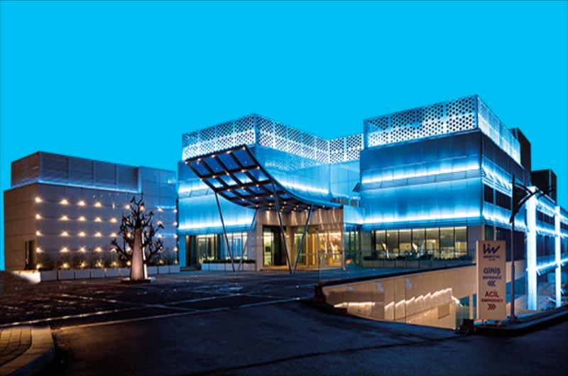
Bayrampaşa Eye Hospital
About Hospital
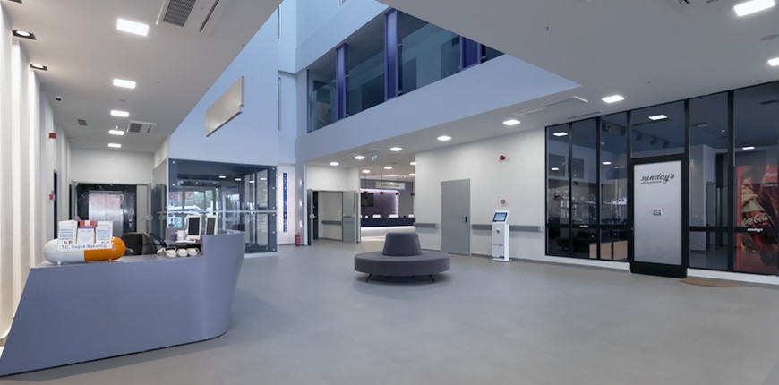
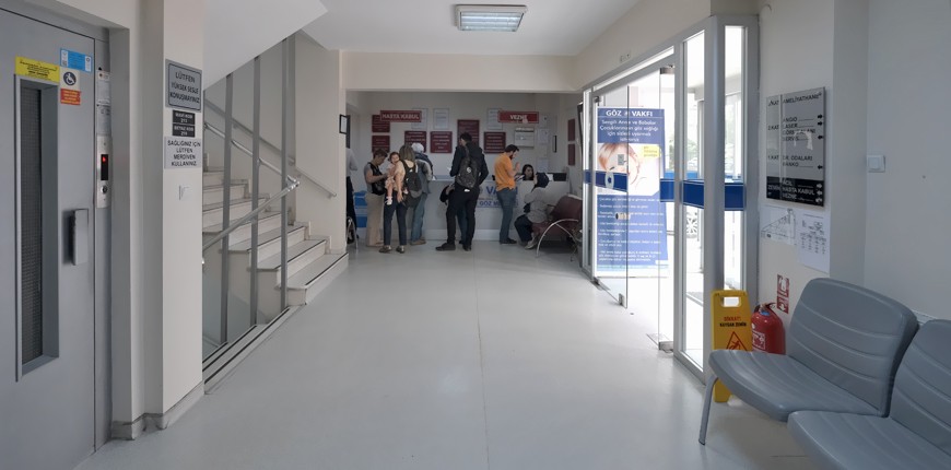
The Foundation was founded by philanthropists in 1984 to eliminate vision defects. The same year, Turkey's first Bayrampaşa Eye Hospital, was opened. In Bayrampaşa Eye Hospital, where all eye health-related treatments and surgeries are performed, services as glaucoma surgery, strabismus, pediatric eye health, cataract, cornea, refractive surgery, oculoplastic surgery and retina units are provided.
Apart from Bayrampaşa Eye Hospital, which is the best in the field of eye, İdealtepe Eye Center (1997) and Yıldırım Eye Center (2001), where surgical operations can be performed, provide all kind of eye health services .
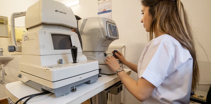
Service buildings, operating rooms, treatment units, and hospitals and eye centers where medical devices are constantly renewed serve with high ethical values and quality.
Bayrampaşa Eye Hospital was established in 1984 purpose and service area of which is to protect the eye health of people "For a Bright Life".
İdealtepe Eye Hospital was opened in 1997 and joined the Eye Foundation family.
Yıldırım Eye Center, which was established in 2001, offers eye health services where surgical operations can be performed.
Bursa Yıldırım Eye Hospital started to serve in 2001.
It is the first Hospital of the Ottoman Empire, built by Yıldırım Beyazıt Han in 1397. It was restored by the General Directorate of Foundations and was put into service by the Foundation in 2001.
The goals of the Foundation:
Early diagnosis
Detection of amblyopia and vision defects in children
Correction and effective treatment
Fight against blindness.
Cataract is when the lens inside the eye loses its transparency and becomes opaque. Surgery is the only known treatment for cataracts, which are most commonly seen due to aging. Cataract surgeries have been performed for nearly 20 years with a technique called phacoemulsification using high ultrasonic waves.
In this surgery, which has an average duration of 20-30 minutes and is performed with drop anesthesia, the lens that has lost its current feature is removed and a new intraocular lens is implanted. Phaco surgery has many stages. The success of the operation may vary depending on the reliability of the operating room environment, the quality of the material used and of course the surgeon's experience.
Femtosecond Laser; It is carried out without human touch, computer controlled.
With the femtosecond phaco operation, the area where the artificial intraocular lens is placed can be properly prepared.
Femtosecond laser and phaco technique;
The eye is anesthetized with drop anesthesia.
After making the necessary adjustments in the femtosecond laser device, the corneal incision, opening in the anterior lens capsule (capsulorhexis) and separating the lens into pieces, which are the important stages of cataract surgery with phaco, are performed completely personally and safely.
Afterwards, patient is taken to another operating room and the processes of removing the lens residues from the eye with Phacoemulsification and placing an artificial intraocular lens are completed.
After these procedures, which are carried out in a period of about 20 minutes, patients can go home.
Excimer laser treatment is a method that has been successfully used in the treatment of refractive errors for more than 20 years. Excimer laser, popularly known as Eye Drawing, is a device that produces 193 nm wavelength ultraviolet light using Ar F gas. The laser beam formed here removes the targeted tissue in the desired thickness and width, so a permanent change occurs in the cornea layer located outside the eye, and the treatment of myopia, hyperopia or astigmatism is realized. Lasik, Femtolasik Lasek and PRK Excimer are different application methods of laser treatment.
LASIK is the most commonly used method in the treatment of focus defects. Patients who are over the age of 18 and have not changed their glasses numbers in the last 1 year are candidates for this treatment. In order to apply Lasik, the refractive error in the eye and the structure of the eye must be appropriate. For this purpose, all candidate patients are subjected to a detailed eye examination as well as a series of examinations showing the characteristics of the corneal layer.
With LASIK, myopia up to 12.0 degrees, hypermetropia up to 6 degrees and astigmatism can be treated. It is important to stop wearing contact lenses before Lasik. It will be appropriate for patients with soft lenses not to use contact lenses for at least one week, and those with hard lenses for at least three weeks.
PRK is the first method used in excimer laser treatment. The epithelium located in the outermost part of the clear cornea layer of the eye is mechanically removed and Excimer laser treatment is applied.
No pain is felt during the procedure.
A contact lens is attached to the patient's eye, and healing occurs within 2 to 3 days. It is a successful method, especially in the treatment of low numbers.
The disadvantage is that pain is felt during the recovery period and the risk of haze (blur) and regression (some regression of the glasses number) is high, especially in the treatment of high numbers.
In LASEK treatment method, the epithelial layer is not removed as in PRK, it is liberated with diluted alcohol solution, and it is placed back after laser treatment is applied.
Recovery is completed after the bandage contact lens application for about 4-5 days.
The epithelial layer left in place acts as a barrier, reducing both postoperative pain and the risk of haze and regression compared to PRK.
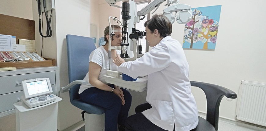
The Retina is the light-detecting layer of the eye. It covers the entire inner wall of the Eye. It consists of vessels and nerves coming from the brain. For retinal examination, after measuring the degree of vision and passing through a microscopic examination, drops that allow the pupil to dilate are dropped. The pupil grows at different times depending on the person and the disease. This period should be waited before the examination. Then, intraocular transparent media (lens and vitreous) and retina are examined in detail with the help of different instruments.
Retinal Detachment
There are complaints such as flashing of light, flying and shadowing in the eye. High myopia and eye blows are the most important risk factors.
If the tear is diagnosed in the early period, its progression is prevented by laser. If there is a delay or negligence in diagnosis and treatment, the tear grows well and the retinal layer becomes detached. In this case, which we call retinal detachment, the only treatment method is surgery.
Macular Degeneration
Until a decade ago, we could not do much to help such patients with treatment. Various treatment methods started to develop after the 2000s. These developments, which started with photodynamic therapy (application of intravenous drugs, laser application), have continued to develop until today with the application of some substances injected into the eye, respectively.
Although the cause of this disease is not clear, heredity and metabolic disorder are thought to be the most important factors. With the examinations to be performed at an early age in the future, people who will have this disease will be identified and treated with gene or other treatment methods before the disease occurs.
ROP
The most important step in the treatment of ROP is regular follow-up. Statistically, 80% of the patients who develop ROP regress spontaneously and treatment is required in 8% of the babies followed up. However, timely determination of the disease level we call threshold disease and urgent (within 3 days) laser treatment are the most important factors that prevent babies who develop ROP from being blind. Vitrectomy surgery is performed in cases that cannot be detected in time and whose disease progresses despite laser treatment.
PDT
Photodynamic therapy; It is applied in some patients with macular degeneration, chronic central serous chorioretinopathy and polypiodal choroidal vasculopathy.
Photodynamic therapy is based on the principle of destroying the target tissue by stimulating this substance with a low-energy light (cold laser) in the target tissue following the intravenous administration of a light-sensitive chemical substance to the body and ensuring the occlusion of the vascular component in the target tissue without damaging the surrounding tissues.
Photodynamic therapy can be repeated if needed. If there is damage in both eyes, it is applied to both eyes simultaneously, if doctor deems appropriate.
Patients under treatment are asked not to be exposed to intense light for 48 hours.
Glaucoma, also known as eye pressure, is an eye disease that occurs with the increase of intraocular pressure and results in blindness if not treated. In the front of the eye, there is a fluid that circulates to nourish the tissues. This liquid is produced inside the eye. Another group is also thrown out of the channel. In some eyes, this fluid cannot be expelled due to the blockage in the ducts and intraocular pressure rises.
The aim in treatment is to protect the patient's vision for life. Glaucoma treatment is in 3 types: medication, laser therapy and surgical treatment.
Treatment is usually started with medication, and if the damage continues, laser or surgical treatment is applied.
Which treatment will be applied depends on the condition of the patient. Early or late diagnosis, age of the patient, and proper use of medications are important factors in the choice of treatment method.
Treatment cannot restore old vision, it only prevents further vision loss.
SLT Laser (Selective Laser Trabeculoplasty) is an alternative treatment option in glaucoma. It provides a supportive solution when treatment is not sufficient in patients using medication. It can only be applied to open angle glaucoma. SLT, which is very effective in correctly selected patients, is a simple treatment method of 3-4 minutes and has no side effects or harm. It can reduce eye pressure by 20-30%. The effectiveness of the treatment, which can be applied every 6 months, continues between 6 months and 2 years.
Keratoconus is a progressive disease that affects both eyes, characterized by the anterior bulging and thinning of the transparent layer (cornea) forming the anterior part of the eye. It is also known as corneal sharpening.
The cornea is the most important eye layer where the rays coming to the eye are refracted. The changes that occur in this area cause the rays to focus incorrectly and seriously impair the quality of vision. The most important complaint in this disease, which is more common in people with family history and allergies, is low vision that cannot be corrected with glasses.
Keratoconus usually starts at the age of 15-16 and can progress up to the age of 35. The course of the disease may vary from person to person. In the early stages of keratoconus, the complaint is usually the frequent change in the number of glasses. As the disease progresses, a clear vision cannot be achieved with glasses.
CCL
It is a method that has been increasingly used for the last 5 years and stops the progression of keratoconus. The structure of the cornea is strengthened by using Ultraviolet A and Riboflavin (Vitamin B2) in the treatment.
Therefore, regardless of the disease level and the visual quality of patients, it is the first treatment option in patients with progress. CCL not only stops the progression of keratoconus, but also provides some increase in the level of vision in some patients.
Cornea Transplant
It is a preferred method in cases where keratoconus progresses and the cornea loses its transparency or when other methods do not provide positive results. Keratoplasty; It is an operation in which the cornea of the eye, which is actually damaged by various diseases, is replaced with the donated cornea, which is known as eye transplant among the people.
In the cornea transplant surgery, a circular piece with a diameter of 6–9 mm is removed from the donated healthy cornea and a piece of the same size is removed from the recipient's (patient's) cornea and sutured to this area. The surgery is preferably done under general anesthesia.
Since keratoconus is a disease seen at a young age, the possibility of developing the same disease in the transplanted cornea or rejection of the newly inserted cornea by the body is high. Therefore, the main goal in keratoconus treatment is to enable patients to continue their lives with their own corneas whenever possible.
- TV in the room
- Medical records transfer
- Interpreter services
- Airport pickup
- Flight booking
- Free Wifi
- Private rooms for patients available
- Parking available
- Nursery / Nanny services
- Visa / Travel office
- Laundry
- Religious facilities
- Rehabilitation
- Personal assistance / Concierge
- Hotel booking
- Local tourism options
- Phone in the room
- Special dietary requests accepted
- Family accommodation
- Spa and wellness
- Beauty salon
- Foreign currency exchange office
- Dry cleaning
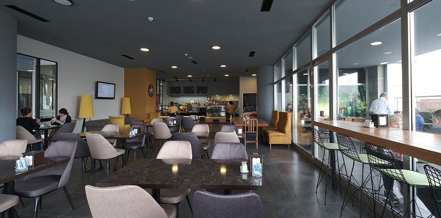
- Restaurante
- Pharmacy
Doctors


Patient Reviews - 0.0
Add Review


-
Hospital and Doctor Search FREE
-
Travel Assistance FREE
-
Transfer Service FREE
-
Translator Service FREE
-
24/7 Online Support FREE