
About Treatment
Electromyography is a diagnostic method that allows you to assess bioelectrical activity of muscles, on the basis of which it can be concluded about the functional state of the nerve that innervates the damaged muscle. This study will help specialist determine the location and extent of the lesion, the severity and nature of muscle and peripheral nerve damage.
Essence of Procedure
This study is carried out using a special apparatus - an electromyograph. Today it is a whole computer system that records the biopotentials of muscles, amplifies them, and then evaluates the data obtained.
Electrodes register muscle potentials and transmit them to an electromyograph. The device amplifies the signal and sends it either to a computer monitor in the form of an image, or to an oscilloscope for subsequent recording on paper.
There are certain norms for the electrical activity of the muscles, indicating their satisfactory function. If the electromyogram readings go beyond these norms, they speak of a disease of the muscle itself or of the peripheral nerve that innervates it.
Types of Electromyography
Depending on the type of electrodes, electromyography is divided into surface (global) and local.
When performing local electromyography, electrode in the form of a thin needle is inserted percutaneously into the thickness of the muscle. This is an invasive technique that is used to study the function of individual muscle elements.
Each type of procedure has its own indications, so the question of which of them should be used is decided individually by the attending physician. Both types of electromyography are often prescribed simultaneously.
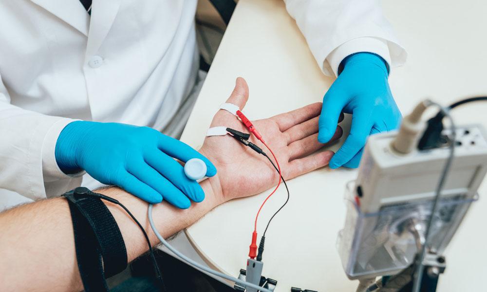
How is Electromyography Performed?
The study can be carried out both in a hospital setting and on an outpatient basis. During it, patient is in a comfortable sitting, half-sitting or lying position. Specialist treats the skin areas that will be in contact with the electrodes with antiseptic and applies electrodes connected to an electromyograph on the muscle to be examined. During the insertion of the needle electrode into the muscle, patient feels a mild pain.
At the beginning of the study, the potentials of the relaxed muscle are recorded, after which patient is asked to slowly strain it and at this time the impulses are also recorded.
The resulting record - electromyogram - is assessed by a specialist in the diagnostic room, and then transmits the conclusion to patient or directly to the attending physician.
Often, electromyography is used in conjunction with a similar study of nerve function - electroneurography. These diagnostic methods complement each other and allow specialist to see the complete picture of a particular disease.
Thus, this study is an important instrumental method for diagnosing a patient's health or disease.
-
Procedure Duration
30-90 minutes
-
Procedure Result
1 hour
Electromyography can be prescribed to a patient if he has the following symptoms or if the following diseases are suspected:
feeling of weakness in the muscles;
frequent intense muscle pain;
frequent muscle twitching, cramps;
Parkinson's disease and syndrome;
ALS (amyotrophic lateral sclerosis);
myoclonus;
myasthenia gravis;
polymyositis;
dystonia;
traumatic damage to peripheral nerves or organs of the central nervous system - the brain or spinal cord;
multiple sclerosis;
botulism;
residual effects after the postponed polymyelitis;
neuropathy of the facial nerve;
tunnel syndromes;
radiculopathy with spinal injuries or hernias of the spinal cord;
polyneuropathy;
essential tremor;
in cosmetology - to determine the areas of the body where botox should be injected.
As a rule, electromyography is performed on the same patient several times. The first examination is at the stage of diagnosis before the start of treatment, and the subsequent ones are in the process of therapy in order to assess its effectiveness.
In general, electromyography is completely safe, harmless and painless study, even for pediatric patients. However, for its implementation, there are contraindications common to many diagnostic procedures:
acute infectious or non-infectious diseases;
EPILEPSY or other organic pathology of the central nervous system;
mental illness, especially those in which patient cannot adequately control himself and perform certain actions;
acute cardiovascular pathology (hypertensive crisis, angina attack, acute stage of myocardial infarction and others);
pacemaker;
skin defects, pustular rashes at the site of the intended impact.
Separately, it should be said about the contraindications to local (needle) electrical stimulation, which are:
if the subject has infections that are transmitted through blood (HIV / AIDS, hepatitis, etc.);
diseases of the blood coagulation system with increased bleeding (hemophilia and others);
high individual pain sensitivity.
Unlike many other diagnostic methods, there are no special preparatory measures for electromyography. However, when planning to go for study, it is worth considering the following points:
stop taking drugs that affect the nervous or muscular system;
several hours before electromyography, do not eat foods that increase anxiety (such as chocolate, coca-cola, tea, coffee, energy drinks).
If you have to take blood-clotting drugs on a daily basis due to a medical condition, be sure to tell your doctor about it.
-
Procedure and Doctor Search FREE
-
Travel Assistance FREE
-
Transfer Service FREE
-
Translator Service FREE
-
24/7 Online Support FREE