About Treatment
Lung cancer is one of the most common cancer type in the world. Men are five times more likely to have lung cancer than women; however, lung cancer cases among women has also increased in recent years. Basically, the disease is directly related to smoking. Most of all, people are sick between the ages of 60 and 70 years.
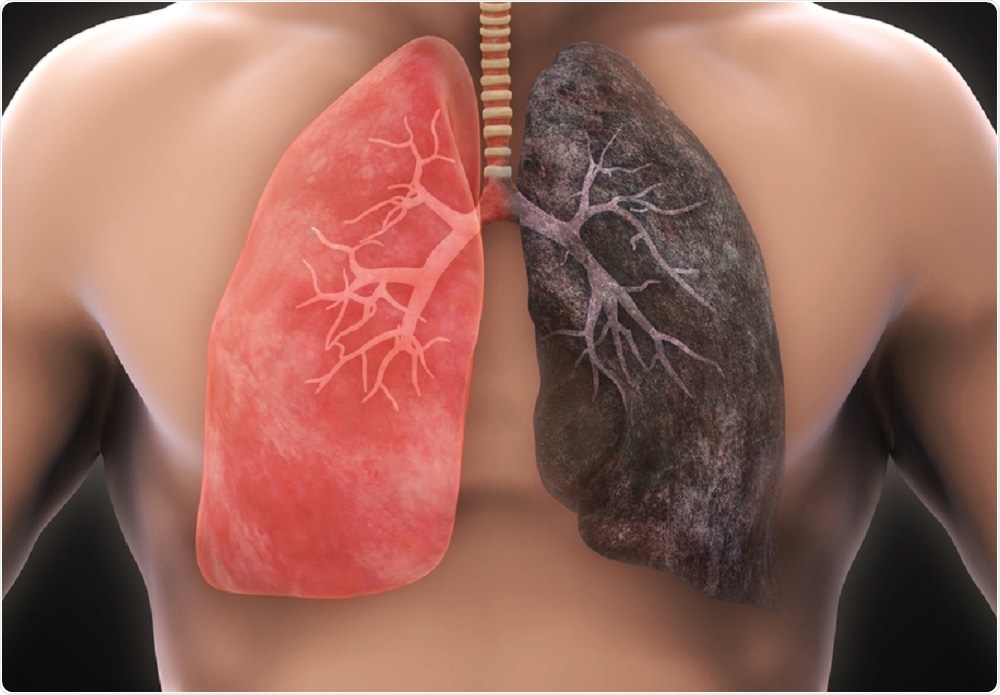
The lungs are soft, spongy, cone-shaped paired organ. The lungs provide respiration - the exchange of carbon dioxide and oxygen. Since the lungs are the internal environment of the body, which constantly comes into contact with the external environment, they have a well-adapted and specialized structure not only for gas exchange, but also for protection - various inhaled infectious pathogens, dust and smoke are retained in the respiratory tract and removed. Three lobes form the right lung, and two lobes on the left. Air enters the lungs through the nasal cavity, throat, larynx and trachea. The trachea is divided into two main bronchi - right and left. The main bronchi are divided into smaller ones and form the bronchial tree. Each branch of this tree is responsible for a small limited part of the lung - a segment. Smaller branches of the bronchi, called bronchioles, pass into the alveoli, in which oxygen and carbon dioxide are exchanged. There are no muscles in the lungs, so they cannot expand and contract on their own, but their structure allows you to follow the respiratory movements that the intercostal muscles and the diaphragm make. The cellular structure of the airways is quite specialized and well adapted for breathing. All airways are lined with epithelium, which is specially adapted cells to perform many important functions as protective; secretion of mucus; removal of irritating substances; onset of immune reactions.
How Lung Cancer Develops?
The formation of lung cancer at the cellular level is not fully understood. Recent studies confirm that the development of lung cancer is not a sudden change in the epithelium of the bronchi. This is a long and complex process, including the gradual development of genetic changes, their accumulation and subsequent cell changes.
Changes in the cells of the bronchial mucosa occur mainly as a result of prolonged exposure to inhaled harmful factors (for example, cigarette smoke, factory pollution). With the gradual accumulation of errors in the mechanisms that regulate cell division, the appearance of cells and their character of division change. The epithelium loses its characteristic appearance, the formation of new cells becomes more and more rapid, and, in the end, control over cell division is generally lost - that is, malignancy occurs. Malignant (cancer) cells are able to multiply and spread throughout the body indefinitely.
At the beginning, lung cancer develops locally and does not grow into the basal (main) membrane of the mucous membrane. With the further development of the process, cancer cells also grow into the basement membrane and penetrate into the underlying connective tissue and vessels. The entire development of the malignant process can last 10 - 20 years and more.
Lung cancer is divided into: small cell lung cancer (SCLC) and non-small cell lung cancer (NSCLC), depending on the type of cancer cells. They differ both in the type of disease course and in the response to the treatment used.
Small cell lung cancer (approximately 15% of all lung cancers) develops from neuroendocrine cells. SCLC is characterized by aggressive growth, often with widespread metastases. Due to its diffuse nature, SCLC is often inoperable, however, it is very sensitive to chemotherapy.
Non-small cell lung cancer is the most common type of lung cancer. It occurs in 85% of lung cancers. Non-small cell lung cancer is further divided:
Adenocarcinoma is the most common type of lung cancer. It covers about 35 - 40% of all cases of lung cancer and its incidence is increasing compared to other types of lung cancer. Adenocarcinoma usually develops on the periphery of the lungs from the mucous glands of the small bronchi. This type of cancer has a tendency to metastasize early to regional lymph nodes and distant sites. Patients with adenocarcinoma may have previously had chronic lung disease, as this cancer often develops in scar tissue.
Squamous cell carcinoma. It takes up about a third of all types of lung cancer. It usually develops in the central part of the airways, therefore, as the tumor grows, the normal air flow is disrupted, shortness of breath and prolonged cough with phlegm develop. There may be hemoptysis. Squamous cell carcinoma is the most common type of lung cancer in smokers.
Large cell carcinoma. It occurs in about 10-15% of all lung cancers. Large cell carcinoma is characterized by rapid growth and widespread metastases. It usually begins in the periphery of the lung and can cause severe chest pain (due to irritation of the pulmonary membrane), as well as accumulation of fluid in the cavity between the lungs and the chest wall, excruciating cough and shortness of breath.
Metastases
Metastases are groups of cancer cells that have moved from the site of cancer to other parts of the body. Metastases can spread along the surfaces of body cavities, lymphatic vessels, or blood vessels.
Bronchocarcinoma can often spread through the lymphatic vessels of the lungs. It can cause severe shortness of breath or, in other words, shortness of breath. The process is usually one-way. The pulmonary lymph nodes are often affected, as well as the lymph nodes in the mediastinum (the space behind the breastbone), axillary and cervical lymph nodes.
Blood from the lungs enters the heart and further into the main blood vessels, which supply blood to all parts of the body. Therefore, metastases in the case of lung cancer can get into virtually any organ, but more often they form in richly vascularized organs - in the bones, liver, adrenal glands, and the brain.
The frequency of metastases depends on the type of malignant cells. For example, distant metastases of small cell lung cancer (SCLC) are much more common than non-small cell lung cancer (NSCLC).
Bone metastases are common complications of lung cancer. They cause severe pain and pathological fractures (i.e., bones break with normal daily stress).
It should be noted that the lungs are the site of metastasis for other tumors, such as renal carcinoma.
Lung Cancer Treatment
Surgery
Radiation therapy
Drug therapy
Lung cancer treatment depends on many factors:
A type of lung cancer (non-small cell or small cell lung cancer);
The size, location and spread of the cancer;
General health of patient.
In turn, patient's quality of life will depend on:
Type of cancer (non-small cell or small cell lung cancer);
Cancer stages;
Treatment regimens.
In patients with progressive disease, the outcome is poor, regardless of the type of pathology, and the goal of treatment is palliative rather than curative. In turn, patients with early diagnosed disease can be treated with various treatments and combinations in the hope of complete cure.
Patients' quality of life is most often affected by cough, respiratory failure, hemoptysis and chest pain. Therefore, the task of treatment is to reduce them as much as possible, while at the same time reducing weakness, improving appetite and emotional state.
Surgery
The decision on whether to have surgery depends on many factors - the location of the tumor, the type and stage of the cancer, and the general health of the patient.
The type of surgery depends on the location of the tumor in the lungs and surrounding tissues. An operation that removes only a small part of the lung is called segmental or wedge resection. If the lobe of the lung is removed, the operation is called lobectomy, and the removal of the entire lung is called pneimonectomy.
Sometimes lung tumor cannot be operated on because of its size or location.
Small cell carcinoma is considered a systemic disease with the risk of widespread dissemination or spread already at an early stage of the disease, so surgical treatment is not successful for most patients. Therefore, each case of SCLC is treated separately, and surgery is not a standard treatment.
In general, only about a third of NSCLC cases can be operated on.
Surgical treatment by stages:
Stage I (T1N0, T2N0) NSCLC is treated with surgical resection whenever possible. The best results are seen in patients with stage T1N0.
Stage II (T1N1, T2N1, T3N0) NSCLC is also treated with surgical resection.
Stage III NSCLC locally or regionally advanced disease that includes large primary tumors (T3 or T4) or mediastinal or supraclavicular (i.e., over the clavicle) lymphadenopathy (i.e., lymph nodes affected by tumor) (N2 or N3) In stage IIIA and IIIB disease (T4 or N3), surgery is always considered, but not always possible.
Radiation Therapy
In the case of lung cancer, radiation therapy is used for therapeutic purposes or to relieve symptoms, since radiation in a short time reduces bone pain, hemoptysis and compression of the superior vena cava syndrome.
Radiation therapy is the local therapy of choice in the case of NSCLC if the tumor is not resectable for objective medical reasons, for example, due to poor lung function.
Stage I and II patients in whom NSCLC is surgically operated, but who have poor lung function, recent myocardial infarction, bleeding tendencies, or other clinical reasons for which surgery must be postponed, may be treated with radiation therapy. The total dose of primary radiation therapy is targeted at the middle of the tumor mass using traditional fractionation or splitting of radiation therapy (30-35 sessions). The duration of the course is approximately 6 - 7 weeks. The goal of therapy is to destroy the primary tumor, all visible pathological lymph nodes and possible microscopic metastases in the lymph nodes.
Patients with stage IIIA NSCLC are rarely treated with just one surgery. In most cases, these patients also receive medication, including chemotherapy and / or radiation therapy: Preoperative chemotherapy (also called neoadjuvant therapy) followed by surgery gives the best results in patients with surgically operated stage IIIA NSCLC. Drug therapy (including chemotherapy) and radiation therapy are standard treatment for patients with stage IIIA NSCLC with good general functional condition, if for some reason surgery is not possible, as well as for patients with stage IIIB NSCLC. Patients with stage IIIB NSCLC with malignant pleural effusion are usually treated as stage IV patients with chemotherapy alone. In patients with stage IV NSCLC, radiation therapy can be used primarily to relieve symptoms.
Radiation therapy can help reduce:
compression of the esophagus (i.e., compression) and swallowing problems,
pain syndrome caused by bone metastases,
neurological symptoms in brain metastases,
compression syndrome of the superior vena cava.
For patients with SCLC, which is localized in only one half of the chest, chemotherapy followed by radiation therapy is considered the standard treatment.
Drug therapy - Chemotherapy
Progressive stages of non-small cell lung cancer are treated mainly with chemotherapy. As with other tumors, chemotherapy with a combination of several drugs is most effective in treating lung cancer.
There is no standard in chemotherapy for non-small cell lung cancer, so different regimens are widely used.
Targeted therapy, monoclonal antibodies
The side effect profile of chemotherapy drugs and drug resistance has prompted scientists to seek new treatment approaches.
EGFR
In lung cancer, excessive production of a special receptor has been proven - the Epithelial Growth Factor Receptor (EGFR). In normal amounts, these receptors provide cellular repair. If there is too much EGFR, it dramatically speeds up the process of cell division until it becomes almost uncontrollable and a tumor forms. The value of EGFR has been proven not only in lung cancer, but also in many other tumors - bowel cancer, head and neck tumors, and pancreatic cancer.
An increased amount of EGFR is associated with an increased risk of metastasis, decreased life expectancy, and poor prognosis.
Currently, new drugs are being used that act directly against the EGFR of cancer cells, preventing them from being activated and, thus, suppressing and limiting tumor growth.
One of the inhibitors of epidermal growth factor is a medication - erlotinib. It inhibits the activity of the enzyme tyrosine kinase, which is required for the EGFR receptor to function, thus interrupting the signaling chain that stimulates the growth and division of cancer cells.
Angiogenesis
For many decades, scientists have been working on the idea of how to not only fight an already formed tumor, but also “not allow” the cancer to grow and develop; prevent the formation of metastases. The efforts were crowned with success - antibodies were created that prevent the formation of blood vessels that supply the cancer.
Both in the tumor and in metastases, an intensive formation of new vessels occurs. Cancer cells themselves "demand" and determine this, by intensely releasing a special blood vessel growth factor (Vascular Endothelial Growth Factor or VEGF). By suppressing the action of this growth factor, the formation of blood vessels decreases and, at the same time, the delivery of nutrients and oxygen to cancer cells decreases. Cancer cells divide rapidly, so their need for oxygen and building material is very high. Thus, the "hunger" caused by antibodies has a destructive effect on cancer; its development is significantly inhibited and complicated. The medication does not work on the mature vessels of a cancerous tumor. The remaining “old” cancer vessels are used to deliver cytotoxic drugs during chemotherapy directly to the tumor and destroy cancer cells. Angiogenesis inhibitors are always used in conjunction with chemotherapy drugs.
-
Types og Lung Cancer
Small cell lung cancer (SCLC) - about 15% of all cases
Non-small cell lung cancer (NSCLC) - about 85% of all cases -
Metastases
Metastases can spread to:
Liver, causing pain, nausea, satiety, and ultimately liver failure
Brain, causing behavior changes, confusion, aphasia, seizures, paresis or paralysis, nausea and vomiting; eventually comes coma and death
Bone, causing severe pain and pathological fractures
Adrenal glands, sometimes causing failure
-Symptoms
-Visual diagnostics
Timely diagnosis of lung cancer is complicated by the fact that there are no specific complaints or signs until cancer has reached a large size. Therefore, in order to make a diagnosis, it is important to evaluate all available information - medical history, exposure to smoking and other harmful factors, symptoms and examination data.
To clarify the causes of symptoms, you need to evaluate:
Medical history - what diseases have been transferred and / or continue to this day;
Smoking - how long has patient been smoking, how many cigarettes a day, etc.
Unfavorable environmental conditions (eg agricultural pollution) and occupationally harmful substances;
Cancer in family members.
The general functional state of patient is a very important prognostic factor, as it determines whether patient will be able to withstand the operation and the rest of the therapy.
When determining the general functional state of patient, patient's malignant diseases and all associated influences, together with other diseases and side diseases, for example, heart or lung diseases, are taken into account. Lung cancer disrupts breathing, and limited breathing over time has a very negative effect on heart activity. Therefore, the general condition of patients with lung cancer is often difficult.
Visual Diagnostics
The main purpose of imaging is to determine the spread of the disease and the stage. Radiography, computed tomography and magnetic resonance imaging are used.
The most commonly used chest X-ray. It is considered the most clinically significant study for the detection of lung cancer, although only a tumor with a size of 1 to 2 cm can be reliably identified. X-ray can detect more than 90% of lung carcinomas.
Computed tomography examination is especially valuable for determining the stage of lung cancer, as it can determine the presence of a tumor in the mediastinum, in the pleura and in the chest wall. A detailed examination of the location and size of the tumor allows you to accurately determine the possibilities of its operation. In addition, it also allows very small tumors and metastases to be identified.
Magnetic resonance (MR) is not used as an everyday routine examination in patients with lung cancer. MRI has more opportunities to determine the structure of nerves and blood vessels, to more accurately assess operable tumors that are close to the lung structures.
Ultrasound examination of the abdominal organs is also used to check and exclude the spread of the disease.
Biopsy
To take a biopsy sample, you can use one of these methods:
* Bronchoscopy - a bronchoscope is inserted through the mouth or nose into the trachea and deeper to examine the airway and obtain small tissue samples
* Aspiration with a needle - a needle is inserted through the chest directly into the tumor and a tissue sample is taken
* Thoracocentesis - a needle is inserted through the chest wall into the pleural cavity to take a sample of the fluid that washes the lungs, in which they are looking for cancer cells
* Thoracoscopy - a thoracoscope (a special tube with a video camera at the end) is inserted through a small incision into the chest cavity and examined as with a magnifying glass.
* Mediastinoscopy - through a small incision over the sternum, a mediastinoscope is inserted into the mediastinum (one of the parts of the chest cavity) and this cavity is examined.
* Thoracotomy is a major operation where the chest is surgically opened.
It is impossible to name any one group of symptoms or specific signs that would indicate lung cancer. However, the most commonly reported lung cancer symptoms can be roughly divided into four broad categories:
local disturbances caused by the growth and spread of cancer;
distant metastases;
non-specific, general symptoms;
paraneoplastic syndromes.
Signs and symptoms that develop in connection with a tumor differ depending on its size and location. For example, centrally located lung cancer tends to cause “squeaking” on breathing, hemoptysis (blood in sputum), shortness of breath (shortness of breath), fever, and persistent cough for no other reason. The spread of lung cancer within the chest (by direct spread or through the lymphatic vessels) is also associated with numerous symptoms and is often the reason why radical (i.e., curing) surgery cannot be performed.
The most striking example of disorders caused by distant metastases are neurological symptoms that cause tumor metastases to the brain: severe headaches, imbalance, decreased sensitivity and / or muscle strength in some part of the body.
In both SCLC and NSCLC, there may be non-specific, systemic signs and symptoms (the so-called carcinomatous intoxication):
loss of appetite,
decrease in body weight,
severe weakness
It should be noted that decreased appetite is often not assessed seriously enough (even in 30% of cases), and is noticed only with a pronounced loss of body weight. General symptoms greatly affect the patient's well-being and quality of life.
There are different syndromes - endocrine, neurological, cardiovascular, skeletal, metabolic (i.e., metabolic) that are considered paraneoplastic.
The general functional state of patient is a very important prognostic factor, as it determines whether patient will be able to withstand the operation and the rest of the therapy.
When determining the general functional state of patient, patient's malignant diseases and all associated influences, together with other diseases and side diseases, for example, heart or lung diseases, are taken into account. Lung cancer disrupts breathing, and limited breathing over time has a very negative effect on heart activity. Therefore, the general condition of patients with lung cancer is often difficult.
-
Procedure and Doctor Search FREE
-
Travel Assistance FREE
-
Transfer Service FREE
-
Translator Service FREE
-
24/7 Online Support FREE