About Treatment
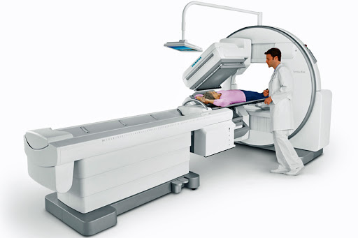
Bone Scintigraphy
Bone scintigraphy (osteoscintigraphy) is a modern screening study for assessing pathological changes in the skeleton with minimal radiation exposure to patient. It should be noted that this method allows you to identify pathological changes in bone tissue before the appearance of radiological signs of the disease.
Three-phase osteoscintigraphy (dynamic 3-stage study) is used mainly for the diagnosis of benign tumors, while the phases of blood flow, tissue blood filling and examination of the bones themselves are assessed.
Most often (mainly with proven malignant neoplasms), the multiphase technique is not required and only the third phase is performed - static scintigraphy of the bones of the whole body.
Purpose of Study
Revealing the localization of zones of benign / malignant disorders of "bone" metabolism.
Metastases of Various Tumors in Bone:
First of all, the suspicion of metastatic lesions of the skeleton arises in breast and prostate cancer, lung cancer, kidney cancer and some others. Particular caution should be exercised with an increase in the level of tumor markers, for example: PSA (prostate-specific antigen), CA 15-3 and some others. After conservative treatment or surgical removal of the tumor, dynamic monitoring of the state of the bone tissue is recommended. Scintigraphy should be performed the first 2 times with an interval of 6-8 months, then, with a normal examination result, after 1–2 years. The need for re-examinations should be clarified with a radiologist or your doctor.
Inflammatory and Traumatic Changes in Bone Tissue:
One of the indications for carrying out radionuclide studies of the bone system (bone scintigraphy) are inflammatory changes in bone tissue. The method allows to determine the prevalence of the process by identifying foci of inflammation in the bones and joints throughout the skeleton, even in the early stages of the disease. On radiographs with osteomyelitis, as a rule, a less prevalence of the process is determined than it actually is. Scintigraphy shows the true size of the inflammatory focus.
Osteoscintigraphy in Orthopedics and Vertebrology:
When prosthetics of joints or the installation of metal structures in the spine, scintigraphy of the bones of the skeleton reveals mechanical instability of the prosthesis components or an inflammatory process around the prosthesis or metal structure. Unlike other research methods (X-ray, CT, MRI), scintigraphy of the bones of the skeleton allows you to determine the intensity of the inflammatory process in different parts of the bone.
-
Contraindications
Pregnancy
-
Duration
1,5-3 hours
Diagnosis, staging, response to therapy and follow-up of tumor (primary tumors - Ewing's sarcoma, osteosarcoma, etc., metastatic lesions as a result of breast, prostate, lung, etc.) and non-tumor diseases (osteomyelitis, Perthes disease, aseptic necrosis, metabolic bone diseases).
Osteoporosis
Fibrous dysplasia
Stress fractures
Joint prosthesis infection
Sacroileitis
Injuries
No special training required. You must have 1 liter of drinking water with you, which must be drunk within 1 hour after the administration of contrast agent . The bladder must be emptied immediately before the examination.
The study is carried out 2-3 hours after the administration of contrast agent. It akes 30 to 45 minutes.
There is a list of drugs that can affect the quality of scintigraphic images of skeletal bones, these include: bicalutamide, estrogens, bisphosphonates, denosumab, corticosteroids, hematopoietic growth factors, iron supplements, methotrexate, nephrotoxic chemotherapy, nifedipine. Therefore, it is necessary to warn radiologist before the study about taking these drugs.
Lack of complex and lengthy preparation for the procedure;
Getting a static or dynamic image of the surveyed area;
Maximum level of safety, since radiation does absolutely no harm to patient;
High information content of results, which makes it possible to accurately assess the degree of damage;
Minimum time for getting the results of the procedure .
During scintigraphy, doctor is able to accurately determine the presence of pathological changes in the body. Most often, scintigraphy is prescribed to detect malignant tumors in bone tissue, find out the true cause of unexplained bone pain, and diagnose complex fractures. In addition, this survey is very often carried out to assess the dynamics of the process of cancer treatment and its effectiveness. You should not be afraid of using a radioactive agent, since its amount introduced into the body for research is so small that it does not cause any side effects.
-
Procedure and Doctor Search FREE
-
Travel Assistance FREE
-
Transfer Service FREE
-
Translator Service FREE
-
24/7 Online Support FREE